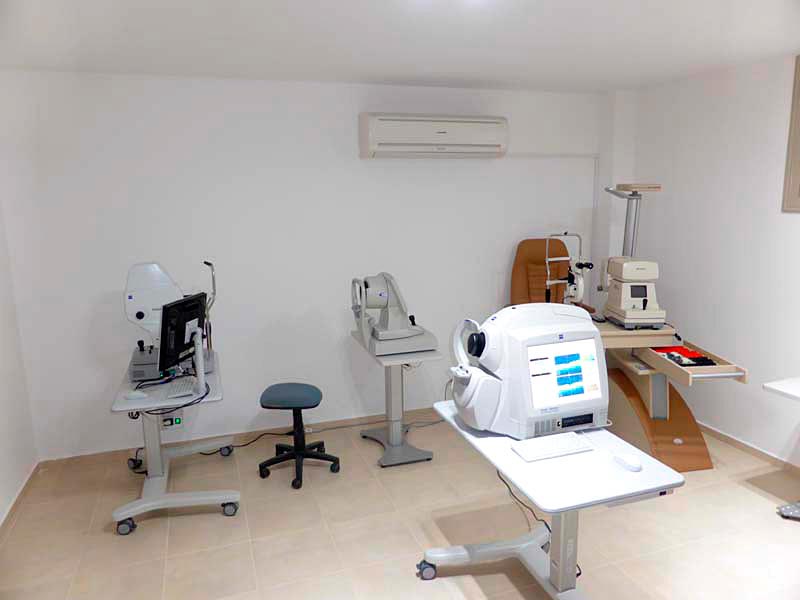
Not many people appreciate their vision as long as they have it and not many people know that the eyes are not just the mirror of our soul but also of our health. Taking the time to visit an eye clinic somebody could find out that with the use of modern equipment and with no machines to touch the body, the eyes can give a glimpse of what is happening inside. At Cult of Vision by the use of the state of the art equipment we can provide you with these kinds of examination which can show not only what is the state of your eyes but also what is the state of your health.
Text: PETER TOKARSKI, CULT OF VISION
OCT examination: 3 D tomography of the Eye
This is the most innovative and comprehensive eye examination which provides the scans of anterior and posterior part of the eye. This scan allows our optometrist to see detailed images of the retina (the innermost layer of the interior eye), enabling him to accurately detect, monitor and control changes to the retina. This procedure is currently the only one that shows in-depth images of the eyes internal structures. Also the optometrist is looking at the cornea images. By analysing the corneal scans it is possible to detect any corneal abnormalities, keratoconus, and corneal thickness at all the corneal layers. Other procedures only show the surface of these structures. An OCT scan can detect the early signs of macular degeneration, glaucoma, detached retinas, keratoconus, corneal dystrophy and other eye disorders. This procedure only takes a few minutes and the equipment never touches the eye, so there is no discomfort.
Retinal pictures
Using the state of the art non-mydriatic fundus camera we can provide you the very detailed examination of the macula, fovea, retina and optic disc. This examimation allows our optometrist to detect all the serious eye diseases like the Glaucoma or AMD. Retinal pictures help also to detect the diabetes and vascular disorders (endocarditis and hypertension).
Corneal Topography
This eye examination allows the optometrist to analyse the corneal abnormalities. It is possible for him to check the surface of cornea and find detailed information about the astigmatism and keratoconus. It also allows designing individual contact lenses which helps to correct high visual disorders. This modern tool helps our optometrist to provide the most satisfied correction of your visual problems.
Slit Lamp examination
The optometrist is performing the evaluation of anterior and posterior parts of the eye using the slit lamp microscope. This eye examination allows the specialist to examine the eye lids, cornea, iris, natural lens and visible part of retina and inform the patient about the health of his eyes.
Vision is probably the most significant out of our five senses and should not be neglected because some of the diseases mentioned can’t be detected with the naked eye. Only with these special examinations and by an expert can be detected and keep always in mind that early diagnosis is half of the cure!
Address: Corner of 25 Martiou and Kostaki Pentalioti, Solomou Square., Paphos, 26955955


